A gain-of-function RAC2 mutation is associated with bone marrow hypoplasia and an autosomal dominant form of severe combined immunodeficiency
Nanolive is happy to introduce a new publication in the Haematologica Journal of The Ferrata Storti Foundation, featuring Nanolive’s 3D Cell Explorer produced by users in the Laboratory of Human Lymphohematopoiesis in Paris, France.
Severe combined immunodeficiencies (SCIDs) are a group of life-threatening genetic disorders that typically present in the first year of life and lead to death if they are not treated quickly. SCIDs are characterized by the absence of autologous T-cells and an intrinsic or functional defect in the B-cell compartment.
Reticular dysgenesis, a severe clinical presentation form of SCID, is characterized by a lack of both neutrophils and T-cells. Individuals suffering from reticular dysgenesis often exhibit bilateral sensorineural deafness in addition.
Molecular diagnosis in SCID patients is key. More than 95% of SCID patients have a molecular diagnosis. All except one one of the known molecular defects in SCID patients involve an autosomal recessive pattern of inheritance (the exception being X-linked inheritance).
Lagresle-Peyrou and colleagues performed biochemical – and in vivo – differentiation assays of patients and human cord blood samples and found a dominant gain-of-function in the RAC2 protein’s GDP/GTP binding site that inhibited hematopoietic stem/progenitor cells differentiation. This gain-of-function mutation leads to a severe autosomal dominant form of SCID with a clinical presentation of reticular dysgenesis. It is the first time that an autosomal dominant mutation has been described as the cause of SCID.
RAC2 gene alterations have been linked to several hematopoietic stem/progenitor cell differentiation-related diseases, leading to the authors to call for RAC2 gene sequencing to be introduced to new-born screening programs for early SCID detection, and for diagnosis of patients suffering from reticular dysgenesis.
Nanolive’s 3D Cell Explorer was used to image patient’s fibroblasts live (acquisition time: 1 image per min over 12 h). The resulting images were then compared to healthy, control fibroblasts in order to determine the growth kinetics of primary fibroblasts. The patient’s sample had a reduced proliferation and increased mortality rate compared to the control. Cell dynamics were also reduced in the patient’s sample, which correlated with the low proliferative response. In addition, cells in the patient’s sample had a highly segmented nuclear membrane and disrupted mitochondrial network compared to control cells.
You can read the full article here!
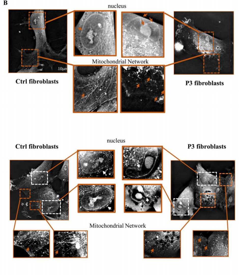
Figure 1. Patient fibroblasts are affected by the G12R mutation. Holotomographic live cell imaging was performed with a 3D Cell Explorer microscope. Cultures of control fibroblasts (left panels) and patient 3’s fibroblasts (right panels) are shown. White arrows indicate the nuclear envelope, and brown arrows indicate the mitochondrial network. Scale bar =10 μm, 1 pixel= 0.188 µm (as in Figure 2B in [1])
References
[1] A gain-of-function RAC2 mutation is associated with bone-marrow hypoplasia and an autosomal dominant form of severe combined immunodeficiency
Chantal Lagresle-Peyrou, Aurélien Olichon, Hanem Sadek, Philippe Roche, Claudine Tardy, Cindy Da Silva, Alexandrine Garrigue, Alain Fischer, Despina Moshous, Yves Collette, Capucine Picard, Jean Laurent Casanova, Isabelle André, Marina Cavazzana
Haematologica Jan 2020, haematol.2019.230250; DOI: 10.3324/haematol.2019.230250
Read our latest news

Revolutionizing lipid droplet analysis: insights from Nanolive’s Smart Lipid Droplet Assay Application Note
Introducing the Smart Lipid Droplet Assay: A breakthrough in label-free lipid droplet analysis Discover the power of Nanolive's Smart Lipid Droplet Assay (SLDA), the first smart digital assay to provide a push-button solution for analyzing lipid droplet dynamics,...

Food additives and gut health: new research from the University of Sydney
The team of Professor Wojciech Chrzanowski in the Sydney Pharmacy School at the University of Sydney have published their findings on the toxic effect of titanium nanoparticles found in food. The paper “Impact of nano-titanium dioxide extracted from food products on...

2023 scientific publications roundup
2023 has been a record year for clients using the Nanolive system in their scientific publications. The number of peer-reviewed publications has continued to increase, and there has been a real growth in groups publishing pre-prints to give a preview of their work....
Nanolive microscopes

3D CELL EXPLORER
Budget-friendly, easy-to-use, compact solution for high quality non-invasive 4D live cell imaging

3D CELL EXPLORER-fluo
Multimodal Complete Solution: combine high quality non-invasive 4D live cell imaging with fluorescence

CX-A
Automated live cell imaging: a unique walk-away solution for long-term live cell imaging of single cells and cell populations