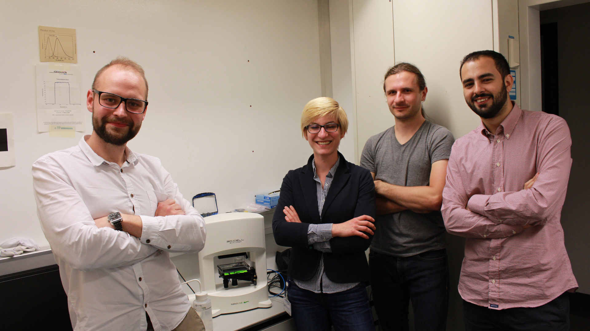
Dr. Yakimovich and colleagues from the Greber group based at the University of Zurich, published a scientific paper that described how Nanolive imaging can be used to visualize the cytopathic (structural changes) effects that different viruses have on host cells during invasion. In the paper, Artur and colleagues compared traditional imaging techniques (phase contrast and fluorescence light microscopy) with those observed using Nanolive imaging and proved that the latter was able to detect the distinct hallmarks of the lifecycle of each infection.
“Nanolive imaging is unique because it is label-free, and cells can be visualized in 3D. So, while we cannot see the viruses themselves, we are able to observe the effect they have on their host cells”. (A. Yakimovich)
Dr. Yakimovich is, like us all, worried about the development of the COVID-19 pandemic, yet he is optimistic that we can defeat it.
“It’s a matter of developing antiviral drugs that are able to treat the infection, but this is easier said than done.”
Scientists have still not been successful for SARS-CoV-2, although there is some hope from previous research on either the SARS or MERS outbreaks. Developing antivirals, he advises me, takes years of painstaking research. First the virus must be isolated, and then grown in highly controlled conditions in a lab and purified until there is enough to study it. You must then understand how the virus penetrates cells, and how it replicates within the human body. Some viruses contain their own (genetic) machinery to replicate, while others hijack the host cells. A great way of understanding the mechanisms involved is via imaging (image-based assays). Traditional imaging methods, such as immunostaining, use fluorescently labelled antibodies to bind to receptors (antigens) on the surface of the virus. While other techniques (such as the addition of GFP-expressing genes) require modification of the genetic code of the virus. Both current approaches are expensive and time consuming. The latter is a big problem for scientists faced by a virus that is spreading so quickly.
Artur is enthusiastic that the approach presented in his paper could shed light on determining how SARS-CoV-2 functions. One of the major advantages of using Nanolive imaging he advises me, “is that you can test the effect of wildtype strains as soon as they have been purified. This could dramatically reduce the amount of time to get results, because it effectively cuts out the need to genetically manipulate the virus beforehand”. I ask whether he has an experiment in mind. “Ideally, I would begin by comparing the responses of wildtype SARS-CoV-2 with the response of other coronaviruses to see if they infect host cells in the same manner in presence or absence of putative inhibitors”.
We encourage scientists working on the topic to get inspired by Artur’s work (https://msphere.asm.org/content/3/6/e00599-18) and if interested in learning more about Nanolive Imaging, get in touch with us (click here).
Read our latest news

Revolutionizing lipid droplet analysis: insights from Nanolive’s Smart Lipid Droplet Assay Application Note
Introducing the Smart Lipid Droplet Assay: A breakthrough in label-free lipid droplet analysis Discover the power of Nanolive's Smart Lipid Droplet Assay (SLDA), the first smart digital assay to provide a push-button solution for analyzing lipid droplet dynamics,...

Food additives and gut health: new research from the University of Sydney
The team of Professor Wojciech Chrzanowski in the Sydney Pharmacy School at the University of Sydney have published their findings on the toxic effect of titanium nanoparticles found in food. The paper “Impact of nano-titanium dioxide extracted from food products on...

2023 scientific publications roundup
2023 has been a record year for clients using the Nanolive system in their scientific publications. The number of peer-reviewed publications has continued to increase, and there has been a real growth in groups publishing pre-prints to give a preview of their work....
Nanolive microscopes

3D CELL EXPLORER
Budget-friendly, easy-to-use, compact solution for high quality non-invasive 4D live cell imaging

3D CELL EXPLORER-fluo
Multimodal Complete Solution: combine high quality non-invasive 4D live cell imaging with fluorescence

CX-A
Automated live cell imaging: a unique walk-away solution for long-term live cell imaging of single cells and cell populations