Automated label-free live cell
imaging & analysis
A revolution for cell biology
eBook – Now available!
“Exploring the frontiers of label-free imaging: Nanolive’s holotomography and AI analysis“ – Download our eBook revealing the complexities of live cell imaging for research and drug development!
Automated label-free live cell
imaging & analysis
A revolution for cell biology
3D Cell Explorer 96focus
Nanolive’s automated label-free live cell imaging and analysis solution
Unlock the power of unlimited high content live cell analysis with the 3D Cell Explorer 96focus. Our unique approach enables analysis and re-analysis of label-free image data with our AI-powered digital assays. As always, the simple, autonomous workflow ensures that your precious time in the lab is used efficiently, and you can be confident that your samples are in good hands, whether you are imaging for an hour, or days.
Streamlined, automated work-flows: save time and money
Long-term monitoring of cells label-free
High content, multiplexed, reliable live cell data
Fully integrated, cutting-edge digital analysis solutions
Fully integrated cutting-edge digital analytical solutions
Get higher significance earlier and faster with our panel of application specific, push-button digital assays. Retroactive analysis is also possible at any time.
Profile cell health and death, and distinguish between living, apoptotic, and necrotic cells label-free.
Measure and characterize simultaneously how live T cells find, bind, stress, and serial kill their targets.
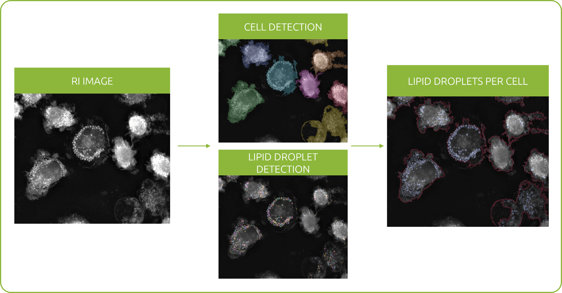
Measure lipid droplet characteristics at population, cellular, and individual droplet levels.
Nanolive imaging and analysis platforms
Swiss high-precision imaging and analysis platforms that look instantly inside label-free living cells in 3D

3D CELL EXPLORER 96focus
Automated label-free live cell imaging and analysis solution: a unique walk-away solution for long-term live cell imaging of single cells and cell populations

3D CELL EXPLORER-fluo
Multimodal Complete Solution: combine high quality non-invasive 4D live cell imaging with fluorescence

3D CELL EXPLORER
Budget-friendly, easy-to-use, compact solution for high quality non-invasive 4D live cell imaging

Latest News


At Nanolive, we focus on innovative product development at the highest standards to deliver a high precision and quality research device for our international customers.