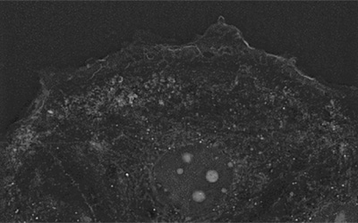
The main event in blood coagulation is fibrin network formation as a result of the polymerization of the soluble plasma protein fibrinogen [1]. Alterations in this process lead to hemostatic and thrombotic disorders such as hemophilia [2] or thrombosis [3]. Additionally, fibrin deposition in tumor cells plays an important role in the formation of the tumor stroma and in angiogenesis [4]. Fibrin clots in cancer exist only within lesions, and their early identification represents a safe and effective method of diagnosis of invasive cancers [4]. The footage shows fibrin network formation in MDA-MB-231 cells, a cell line from human breast adenocarcinoma. An image was obtained every 20 seconds during 15 minutes on Nanolive’s 3D Cell Explorer.
[1] Palta, S., Saroa, R., & Palta, A. (2014). Overview of the coagulation system. Indian journal of anaesthesia, 58(5), 515–523. https://doi.org/10.4103/0019-5049.144643
[2] Brummel-Ziedins, K. E., Branda, R. F., Butenas, S., & Mann, K. G. (2009). Discordant fibrin formation in hemophilia. Journal of thrombosis and haemostasis : JTH, 7(5), 825–832. https://doi.org/10.1111/j.1538-7836.2009.03306.x
[3] Korte W, Poon M, -C, Iorio A, Makris M: Thrombosis in Inherited Fibrinogen Disorders. Transfus Med Hemother 2017;44:70-76. doi: 10.1159/000452864
[4] Obonai, T., Fuchigami, H., Furuya, F. et al. Tumour imaging by the detection of fibrin clots in tumour stroma using an anti-fibrin Fab fragment. Sci Rep 6, 23613 (2016). https://doi.org/10.1038/srep23613
Read our latest news
Revolutionizing lipid droplet analysis: insights from Nanolive’s Smart Lipid Droplet Assay Application Note
Introducing the Smart Lipid Droplet Assay: A breakthrough in label-free lipid droplet analysis Discover the power of Nanolive's Smart Lipid Droplet Assay (SLDA), the first smart digital assay to provide a push-button solution for analyzing lipid droplet dynamics,...
Food additives and gut health: new research from the University of Sydney
The team of Professor Wojciech Chrzanowski in the Sydney Pharmacy School at the University of Sydney have published their findings on the toxic effect of titanium nanoparticles found in food. The paper “Impact of nano-titanium dioxide extracted from food products on...
2023 scientific publications roundup
2023 has been a record year for clients using the Nanolive system in their scientific publications. The number of peer-reviewed publications has continued to increase, and there has been a real growth in groups publishing pre-prints to give a preview of their work....
Nanolive microscopes
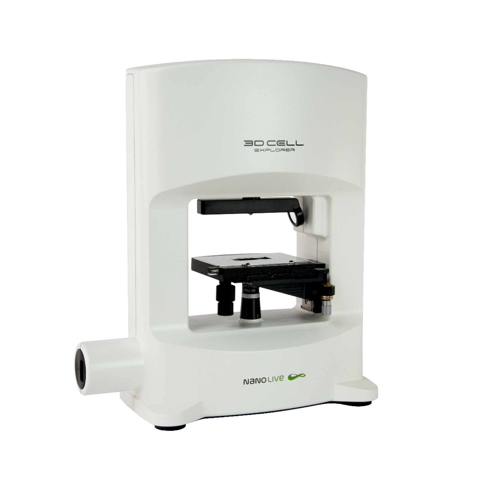
3D CELL EXPLORER
Budget-friendly, easy-to-use, compact solution for high quality non-invasive 4D live cell imaging
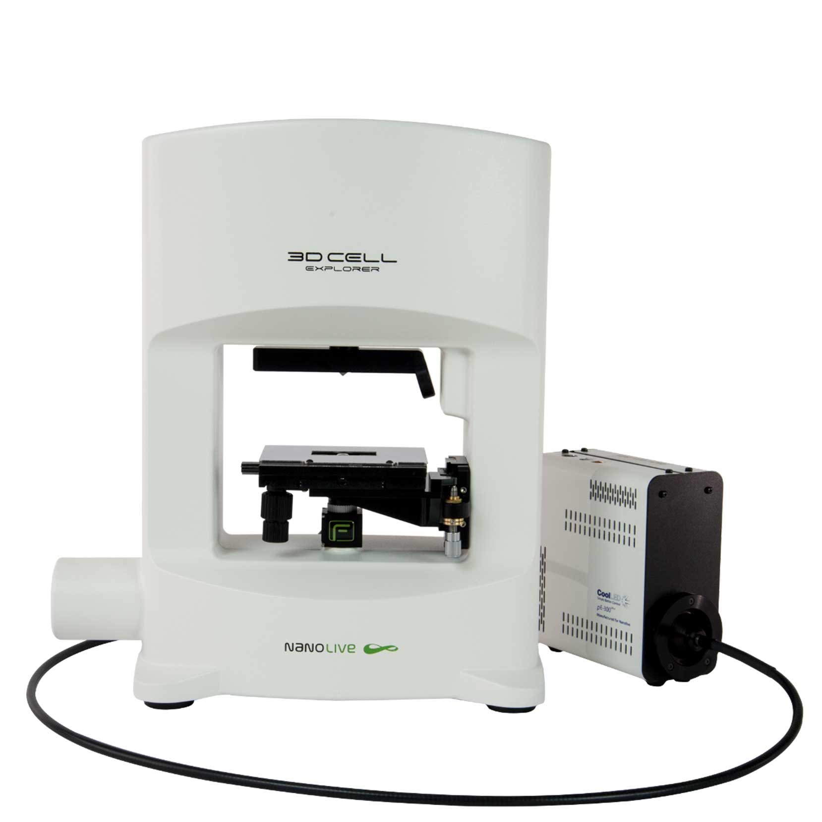
3D CELL EXPLORER-fluo
Multimodal Complete Solution: combine high quality non-invasive 4D live cell imaging with fluorescence
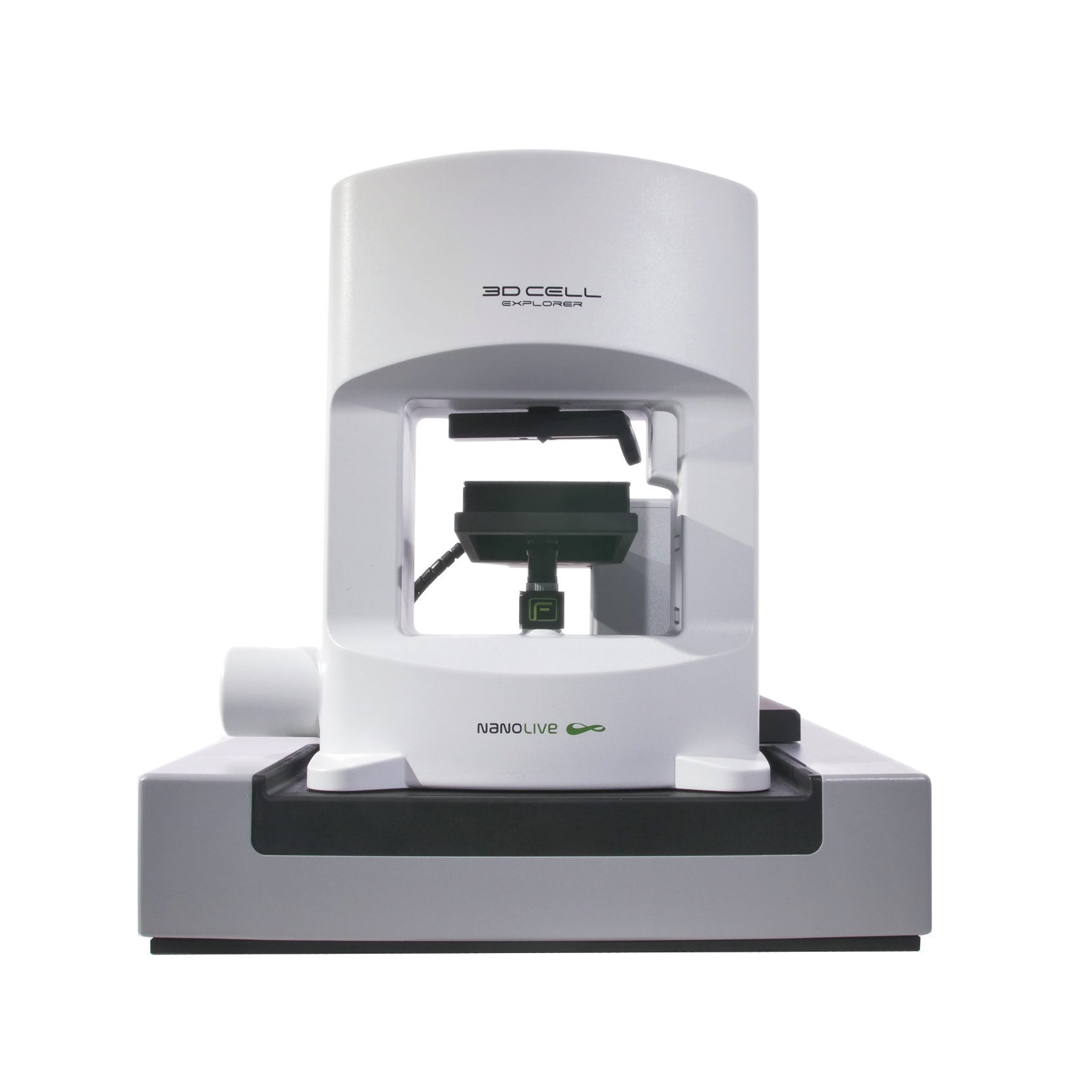
CX-A
Automated live cell imaging: a unique walk-away solution for long-term live cell imaging of single cells and cell populations

