It is almost impossible to image the inside of a living cell without damaging it, even with the most modern devices. Traditional microscopy techniques require researchers to prepare their samples well in advance (1–72 hours). These procedures are likely to be invasive to the cells (risk of cell damage and cell stress). The 3D Cell Explorer was created using a disruptive technology which, for the first time, allows users to explore instantly the inside of a living cell in 3D without the need for any labeling or other invasive methods.
Similar to computer tomography for human bodies
Just like computer tomography (CT) for human bodies, the 3D Cell Explorer can make a complete tomographic data set of the living cell. This happens instantly and without any harm to the live cell.
The 3D Cell Explorer’s technology is unique worldwide. It allows determining how light propagates through the cell. By this means, one can measure the cell’s physical properties, i. e. the refractive index (RI). The result is quantitative cell tomography, in vitro without any invasion or sample preparation.

Holotomography – a revolutionary technology
The combination of holography and rotational scanning makes Nanolive a revolutionary technology.
Holography offers a unique means to measure cells in their native environment: label-free, non-invasive, manipulation-free, and interference-free. Rotational scanning allows 3D reconstructions, noise robustness, and a resolution far beyond the accepted limit for light.
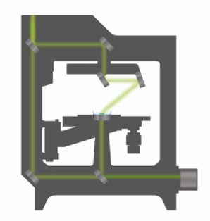
The sample is positioned between a high-numerical-aperture air objective beneath the sample and a rotational illumination arm above. Green light (520 nm) from a laser diode is split into sample and reference beam. The sample is illuminated with a laser beam inclined at 45° which rotates around the sample 360°. A series of holograms is recorded on a digital camera by combining the beam that has passed through the sample with the reference beam.
Applications
Non-invasive 3D characterization of live cells in physiological conditions: With the 3D Cell Explorer it is possible to measure the quantitative refractive index of cell organelles in seconds. Hence, it is possible to do non-invasive in vitro imaging of almost any kind of cells with up to 30 μm depth of reconstruction. This allows for biological features to be segmented based on their physical characteristics.

Label-free 4D continuous observation of cell processes from seconds to weeks: The 3D Cell Explorer enables to study cell life cycle processes of growth, division & death in 3D and 4D. With a dedicated top-stage incubator, cell compartments and their kinetics and dynamics in real-time at every second without interfering with their natural physiology can be monitored.

Multiplexing: High resolution and high sensitivity characterization of multiple cell organelles based on their refractive index. the 3D Cell Explorer makes it possible to explore and measure up to 8 cell organelles simultaneously with unprecedented detail and resolution, marker-free and preparation-free based on their own physical density.

Visit www.nanolive.ch for more information.