This video shows the versatility of the 3D Cell Explorer microscope in imaging both very fast and very slow processes without compromising with cell health and at the desired frequency.
There are 4 different cell types. Can you guess the name of each cell death when reading the explanations?
Video A: Necrosis
This is a very rapid process characterized by swelling of organelles, dilatation of the nuclear membrane, increased cell volume (oncosis), culminating in the disruption of the plasma membrane and subsequent loss of intracellular contents. ID8-ova cells (ID8 murine ovarian tumor cell line transduced with ovalbumin). NaOh was added to the medium during the acquisition to trigger this type of cell death. The cells were imaged for 2 minutes at a frequency of 1 image every 2 seconds.
Video B: Pyroptosis
In this video mouse macrophages were infected with Listeria. The immune cells recognize the foreign danger signals within themselves, release pro-inflammatory cytokines, swell, burst and die. These cells were imaged for 25 minutes at a frequency of 1 image every 15 seconds.
Video C: Apoptosis
This is a Programmed Cell Death of T685A human melanoma cancer cell. During real-time monitoring, we can observe the typical morphological cellular changes as blebbing, cell shrinkage and chromatin condensation. The cells were imaged for 2h30 at a frequency of 1 image every 30 seconds.
Video D Autophagy
This cell death consists of the formation of a double membrane vesicle around targeted cellular components. This vesicle, known as an autophagosome, then migrates through the cytosol to merge with a lysosome filled with hydrolytic enzymes, resulting in the breakdown of the autophagosomes contents. Cell death was induced by the addition of a drug (combination of imipramine and ticlopidine) to mouse glioblastoma cells (LN18). The cells were imaged every 60 seconds over a period of 9 hours. Special thanks to the Laboratory of Translational Oncology – Prof. Douglas Hanahan from EPFL.
Simultaneous, massive mammalian cell necrosis
Example of a co-culture experiment performed using Nanolive’s 3D Cell Explorer. Mouse skin melanoma cancer cells (B16) were incubated overnight with Dictyostelium amoebae cells (WT1), in order to visualize their interactions.
We observed a simultaneous, massive mammalian cell necrosis, the reason of which remains unknown. What do you think is happening here? Why do these cells suddenly explode?
Experimental conditions: Mouse skin melanoma cancer cells (B16, p35) were grown to 40% confluency in complete DMEM medium. Dictyostelium amoebae cells (WT1, p14) were previously grown in HL-5 medium. The time-lapse imaging experiment was conducted with a standard top-stage incubator set to 37°C and 5% CO2 for 8 hours, capturing images every minute.
Read our latest news
Revolutionizing lipid droplet analysis: insights from Nanolive’s Smart Lipid Droplet Assay Application Note
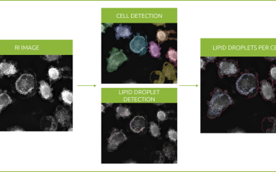
Introducing the Smart Lipid Droplet Assay: A breakthrough in label-free lipid droplet analysis Discover the power of Nanolive's Smart Lipid Droplet Assay (SLDA), the first smart digital assay to provide a push-button solution for analyzing lipid droplet dynamics,...
Food additives and gut health: new research from the University of Sydney
The team of Professor Wojciech Chrzanowski in the Sydney Pharmacy School at the University of Sydney have published their findings on the toxic effect of titanium nanoparticles found in food. The paper “Impact of nano-titanium dioxide extracted from food products on...
2023 scientific publications roundup
2023 has been a record year for clients using the Nanolive system in their scientific publications. The number of peer-reviewed publications has continued to increase, and there has been a real growth in groups publishing pre-prints to give a preview of their work....


